
What is an EKG?
An EKG, also known as an ECG, stands for Electrocardiogram—a diagnostic tool that measures and records the electrical activity of the heart over a period of time. Through the use of small electrode patches attached to the skin, the EKG captures the heart's rhythmic electrical impulses and translates them into wavy lines on a graph paper or digital screen. These tracings offer a visual representation of the heart's beat pattern, highlighting any irregularities or disturbances in its normal rhythm. EKGs have become an integral part of medical diagnostics because they can provide crucial insights into the overall health of the heart, assist in detecting various cardiac conditions like arrhythmias, and guide treatment decisions. In essence, an EKG is a window into the heart's electrical functionality, giving doctors a real-time snapshot of its performance and potential issues.

The Human Heart’s Electrical System
The heart, often thought of as merely a muscular pump, is also an intricately designed electrical system. This system is responsible for initiating and coordinating the heartbeat. At its core is a group of unique cells known as the sinoatrial (SA) node, located in the right atrium. Often referred to as the heart's natural pacemaker, the SA node fires electrical impulses that trigger each heartbeat. These impulses then travel through a pathway of specialized cells, causing both the atria and the ventricles to contract in a synchronized manner, ensuring efficient blood circulation. Following the SA node, the electrical impulse moves to the atrioventricular (AV) node, then down the bundle of His, and finally through the Purkinje fibers, coordinating the heartbeat from the atria to the ventricles. This coordinated electrical activity not only drives the heart's rhythmic contractions but also ensures that blood is pumped efficiently to nourish every cell in the body. Any disruption in this electrical sequence can lead to arrhythmias or irregular heart rhythms, highlighting the importance of this system in maintaining cardiovascular health.
Basic Components of an EKG Machine
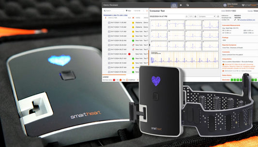
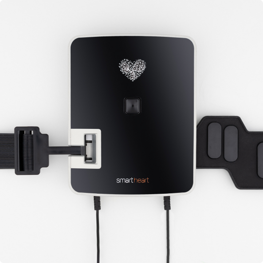
An EKG machine is a sophisticated device designed to capture the intricate electrical activities of the heart, and it comprises several essential components that ensure its accuracy and functionality. First and foremost are the electrodes, small adhesive patches that are strategically placed on the patient's skin, typically on the chest, arms, and legs. These electrodes detect the heart's electrical impulses. Connected to these electrodes are the lead wires, which serve as the conduits for transmitting the detected electrical signals to the main EKG machine. The main machine, often a combination of hardware and software, processes these signals and displays them as tracings on graph paper or a digital screen. This display provides a visual representation of the heart's electrical activity. Within the machine, amplifiers magnify the signals, filters refine them for clarity, and a recording device (either digital or paper-based) captures the final EKG reading. Together, these components work seamlessly to give medical professionals a real-time snapshot of the heart's electrical rhythm and potential anomalies therein.
The Role of Electrodes
Electrodes play a pivotal role in the EKG process, acting as the primary interface between the patient's body and the EKG machine. These small, round patches, often made of a conductive material like silver or silver chloride, are strategically adhered to the skin to detect the minute electrical impulses produced by the heart. Once an impulse is detected, it is the electrode's responsibility to transmit this signal, with minimal interference, to the EKG machine via the lead wires. There are different types of electrodes, including adhesive patches for single-use and suction or clamp electrodes for repeated uses. Some are designed specifically for areas like the chest, known as precordial electrodes, while others, called limb electrodes, are intended for placement on the arms and legs. The precise placement and quality of these electrodes are paramount, as they directly influence the clarity and accuracy of the EKG reading. Misplacements or faulty electrodes can lead to artifacts or erroneous readings, underscoring their vital role in obtaining a clear, interpretable picture of the heart's electrical activity.
The Science Behind the EKG Tracings
EKG tracings, with their characteristic wavy lines, are a visual representation of the heart's electrical journey, captured in real-time. As the heart's electrical impulses are picked up by the electrodes, they produce a voltage that varies over time, corresponding to the different phases of the heart's rhythm. The EKG machine measures these voltage changes relative to time. When the heart's chambers are at rest, the line is flat, but when an electrical impulse passes through, it creates a deviation from this baseline. This deviation is what we observe as the ups and downs on the EKG tracing. Each significant wave or segment on the EKG—be it the P wave, the QRS complex, or the T wave—corresponds to specific electrical activities and mechanical contractions of the heart. For instance, the P wave represents atrial depolarization, while the QRS complex depicts ventricular depolarization. The entire tracing essentially maps out the heart's electrical cycle, from the initiation of an impulse in the sinoatrial node to the completion of a heartbeat. By analyzing these tracings, clinicians can discern not only the rhythm and rate of the heart but also detect irregularities or abnormalities that may suggest underlying cardiac conditions.
Safety Aspects of an EKG Test
An EKG test is one of the safest diagnostic procedures in the realm of cardiology. The machine doesn't emit electricity but merely records the heart's natural electrical activity, ensuring there's no risk of electrical shock to the patient. The electrodes used are passive; they listen and convey the heart's signals without introducing any external electrical currents. Some patients might worry about potential discomfort, but rest assured, the adhesive patches used to attach the electrodes are designed to be skin-friendly, and while there might be a brief cold or tingling sensation when the electrodes are placed, the test itself is painless. Additionally, there's no radiation involved in an EKG, unlike some other diagnostic tests, so patients aren't exposed to any radiation risks. For those concerned about allergic reactions, it's worth noting that allergic reactions to the electrode adhesive are rare. However, if any skin irritation does occur, it's typically mild and resolves quickly once the electrodes are removed. Overall, the EKG test's non-invasive nature, combined with its passive recording approach, makes it a safe and essential tool in cardiac diagnostics.
In Conclusion
Understanding the inner workings and significance of the EKG heart monitor offers a fascinating glimpse into the world of cardiology and medical diagnostics. This incredible tool, with its electrodes, intricate tracings, and advanced machinery, bridges the gap between the palpable heartbeats we feel and the complex electrical symphony that orchestrates them. While technology continues to evolve, the fundamental goal remains consistent: ensuring heart health and providing timely, accurate insights to medical professionals. An EKG is not just a testament to human innovation but also a powerful reminder of the heart's intricate design and the crucial role it plays in our overall well-being. Rest assured, with the safety and precision inherent in this procedure, the EKG will continue to be an invaluable cornerstone in cardiac care for years to come.